E-sayfa Özgün Görüntüler E-page Original Images E
advertisement

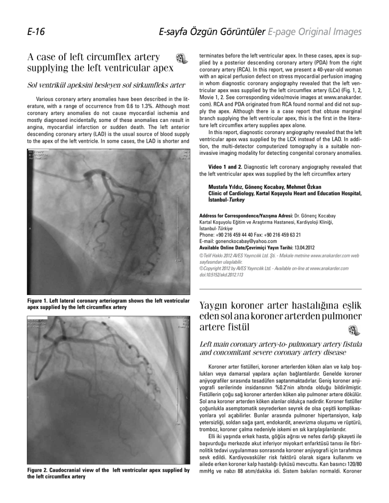
E-16 E-sayfa Özgün Görüntüler E-page Original Images A case of left circumflex artery supplying the left ventricular apex Sol ventrikül apeksini besleyen sol sirkumfleks arter Various coronary artery anomalies have been described in the literature, with a range of occurrence from 0.6 to 1.3%. Although most coronary artery anomalies do not cause myocardial ischemia and mostly diagnosed incidentally, some of these anomalies can result in angina, myocardial infarction or sudden death. The left anterior descending coronary artery (LAD) is the usual source of blood supply to the apex of the left ventricle. In some cases, the LAD is shorter and terminates before the left ventricular apex. In these cases, apex is supplied by a posterior descending coronary artery (PDA) from the right coronary artery (RCA). In this report, we present a 40-year-old woman with an apical perfusion defect on stress myocardial perfusion imaging in whom diagnostic coronary angiography revealed that the left ventricular apex was supplied by the left circumflex artery (LCx) (Fig. 1, 2, Movie 1, 2. See corresponding video/movie images at www.anakarder. com). RCA and PDA originated from RCA found normal and did not supply the apex. Although there is a case report that obtuse marginal branch supplying the left ventricular apex, this is the first in the literature left circumflex artery supplies apex alone. In this report, diagnostic coronary angiography revealed that the left ventricular apex was supplied by the LCX instead of the LAD. In addition, the multi-detector computerized tomography is a suitable noninvasive imaging modality for detecting congenital coronary anomalies. Video 1 and 2. Diagnostic left coronary angiography revealed that the left ventricular apex was supplied by the left circumflex artery Mustafa Yıldız, Gönenç Kocabay, Mehmet Özkan Clinic of Cardiology, Kartal Koşuyolu Heart and Education Hospital, İstanbul-Turkey Address for Correspondence/Yaz›şma Adresi: Dr. Gönenç Kocabay Kartal Koşuyolu Eğitim ve Araştırma Hastanesi, Kardiyoloji Kliniği, İstanbul-Türkiye Phone: +90 216 459 44 40 Fax: +90 216 459 63 21 E-mail: gonenckocabay@yahoo.com Available Online Date/Çevrimiçi Yayın Tarihi: 13.04.2012 ©Telif Hakk› 2012 AVES Yay›nc›l›k Ltd. Şti. - Makale metnine www.anakarder.com web sayfas›ndan ulaş›labilir. ©Copyright 2012 by AVES Yay›nc›l›k Ltd. - Available on-line at www.anakarder.com doi:10.5152/akd.2012.113 Figure 1. Left lateral coronary arteriogram shows the left ventricular apex supplied by the left circumflex artery Yaygın koroner arter hastalığına eşlik eden sol ana koroner arterden pulmoner artere fistül Left main coronary artery-to- pulmonary artery fistula and concomitant severe coronary artery disease Figure 2. Caudocranial view of the left ventricular apex supplied by the left circumflex artery Koroner arter fistülleri, koroner arterlerden köken alan ve kalp boşlukları veya damarsal yapılara açılan bağlantılardır. Genelde koroner anjiyografiler sırasında tesadüfen saptanmaktadırlar. Geniş koroner anjiyografi serilerinde insidansının %0.2’nin altında olduğu bildirilmiştir. Fistüllerin çoğu sağ koroner arterden köken alıp pulmoner artere dökülür. Sol ana koroner arterden köken alanlar oldukça nadirdir. Koroner fistüller çoğunlukla asemptomatik seyrederken seyrek de olsa çeşitli komplikasyonlara yol açabilirler. Bunlar arasında pulmoner hipertansiyon, kalp yetersizliği, soldan sağa şant, endokardit, anevrizma oluşumu ve rüptürü, tromboz, koroner çalma nedeniyle iskemi en sık karşılaşılanlarıdır. Elli iki yaşında erkek hasta, göğüs ağrısı ve nefes darlığı şikayeti ile başvurduğu merkezde akut inferiyor miyokart enfarktüsü tanısı ile fibrinolitik tedavi uygulanması sonrasında koroner anjiyografi için tarafımıza sevk edildi. Kardiyovasküler risk faktörü olarak sigara kullanımı ve ailede erken koroner kalp hastalığı öyküsü mevcuttu. Kan basıncı 120/80 mmHg ve nabzı 88 atım/dakika idi. Sistem bakıları normaldi. Koroner