Weighting and planes
T2-weighted axial
Field-ofview
TR
TE
Sequence
115
5320
100
Turbo Spin Echo
Slice
GAP
Matrix size
thicknessRadyoloji Özel Sayısı
3.2 mm
0.3 mm
512 × 512
Tablo 1: MR görüntüleme tekniği
Proton Density-weighted
Ağırlıklandırma ve
axial fat
suppressed
düzlemler
Görüş
150
aksiyel
ProtonT2-ağırlıklı
Density-weighted
sagittal
140
Yağ süpresyonlu proton
yoğunluk ağırlıklı aksiyel
Proton Density-weighted
sagittal fat suppressed
Proton yoğunluk ağırlıklı
sagital
Proton Density-weighted
coronal
Yağ süpresyonlu proton
yoğunluk ağırlıklı sagital
Proton Density-weighted
Proton
yoğunluk ağırlıklı
coronal
fat suppressed
koronal
Yağ süpresyonlu proton
yoğunluk ağırlıklı koronal
TR
1200
TE45
alanı
Turbo Spin Echo fat
Sekans
Kesit 2 mmBoşluk 0.0 mm
Matris 256 × 256
suppressed
ebatları
kalınlığı
115
5320
100
Turbo Spin Echo
1200
45
Yağ süpresyonlu
Turbo Spin Echo
2570
150
150
1200
140
2570
120
2770
150
1200
160
6040
120
160
41
45
41
31
45
41
2770
31
6040
41
Turbo Spin Echo
3,2 mm
0,3 mm
2 mm
0,0 mm
2 mm
Turbo Spin Echo
fat suppressed
Turbo Spin Echo
2 mm
2 mm
Turbo Spin Echo
Yağ süpresyonlu
Turbo Spin Echo
0,0 mm
256 × 256
256x256
0.3 mm
0,0 mm
256 × 256
256x256
0.0 mm
3 mm
2 mm
512x512
0.0 mm
640 × 640
256x256
Turbo Spin Echo
2 mm
0.3 mm
512 × 512
fatSpin
suppressed
Turbo
Echo
3 mm
0,3 mm
640x640
Yağ süpresyonlu
Turbo Spin Echo
2 mm
0,3 mm
512x512
Case 2: Melorheostosis
Vaka 2: Meloreostozis
Patient
history
Hastanın hikayesi
25-year-old
maleözellikle
with right
knee
pain,
Sağ dizinde,
patellanın
mediyal
ve
posterior
sınırı
boyunca
ağrısı
olan
25
mainly along the medial border of his
yaşında
erkek
hasta.
Hasta,
2006
yılında
sağ
patella and posterior. He states he hyperdizinde hiperekstansiyon olduğunu söyledi.
extended his right knee in 2006. He
O dönemde değerlendirmeye tabi tutulan
was evaluated
and was diagnosed with
hastaya, meloreostozis teşhisi konmuş,
melorheostosis
and
possible
fracture
patellada olası
bir akırık
ve hareketli
gevşek
of theyapılar
patella
with
migrating
loose
tespit
edilmiş.
Konservatif
bir bodşekilde
tedavi
hasta,
o tarihten beri ağrısının
ies and
wasedilen
treated
conservatively.
He
kalıcı
hale
geldiğini
belirtti;
ayrıca
dizinde
states that his pain has been continuous
zaman He
zaman
kilitlenme,
tutulma,
çözülme,
since then.
notes
locking,
catching,
tıklama ve sıkışma olduğunu ekledi.
giving way, popping and grinding.
Fiziki muayene
Physical
examination
Sağ dizin fokal analizinde herhangi bir
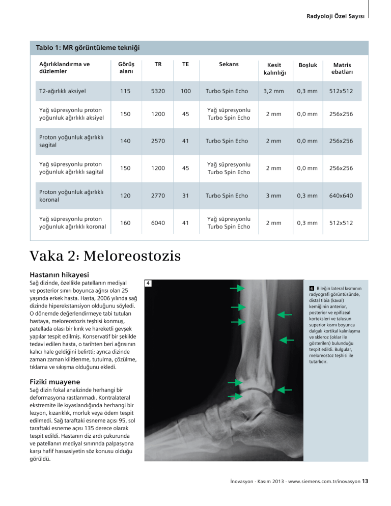
4
44 Bileğin
lateral
kısmının
Lateral
ankle
radyografi
görüntüsünde,
radiograph
showdistal tibia (kaval)
ing undulating
kemiğinin anterior,
cortical thickening
posterior ve epifizeal
and sclerosis
korteksleri
ve talusun
(arrows)
along
the
superior
kısmı
boyunca
dalgalı
kortikal
kalınlaşma
anterior,
posterior,
ve
skleroz
(oklar ile
and
epiphyseal
gösterilen) bulunduğu
cortices of the distespit edildi. Bulgular,
tal tibia, as
wellile
meloreostoz
teşhisi
as involving the
tutarlıdır.
superior talus.
Findings are consistent with
melorheostosis.
Focal exam
of rightrastlanmadı.
knee reveals
no defordeformasyona
Kontralateral
ekstremite
ile
kıyaslandığında
herhangi
bir
mity. No lesions, erythema, ecchymosis
lezyon,
kızarıklık,
morluk
veya
ödem
tespit
or edema when compared to the contraSağ taraftaki esneme açısı 95, sol
lateraledilmedi.
limb. Flexion
on his right 95 and
taraftaki esneme açısı 135 derece olarak
on his left is 135 degrees. He has slight
tespit edildi. Hastanın diz ardı çukurunda
tenderness
to palpation
in the popliteal
ve patellanın
mediyal sınırında
palpasyona
fossa as
well
medial border
of his
karşı
hafifashassasiyetin
söz konusu
olduğu
görüldü.
patella.
İnovasyonFlash
· Kasım
2013 · www.siemens.com.tr/inovasyon
13
MAGNETOM
· 2/2012
· www.siemens.com/magnetom-world

